ISBN:
9780323327459
Copyright:
2016
Publication Date:
05-28-2015
Page Count:
464
Imprint:
Elsevier
List Price:
$53.99

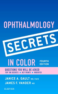
Ophthalmology Secrets in Color Elsevier eBook on VitalSource, 4th Edition
by Janice Gault, MD, FACS and James F. Vander, MD
Elsevier eBook on VitalSource
ISBN:
9780323327459
Copyright:
2016
Publication Date:
05-28-2015
Page Count:
464
Imprint:
Elsevier
List Price:
$53.99
Was
$53.99
Now $37.79
Or $0.00 with a valid access code
Portable and high yield, Ophthalmology Secrets in Color, 4th Edition is perfect for use in clerkships, for exam prep, or as a handy clinical reference. From basic science to problems of the aging eye, it covers everything you need to stay abreast of the latest in this broad specialty. A bestselling volume in the popular Secret Series®, its Q&A format, helpful lists and tables, and informal tone combine to make reference fast, easy, and enjoyable.
New Edition Expected Availability: 02-10-2022

Ophthalmology Secrets Elsevier eBook on VitalSource
Elsevier eBook on VitalSource
ISBN: 9780323674690
-
- A question-and-answer format, bulleted lists, mnemonics, and practical tips from the authors expedite reference and review
- Two-color page layout, "Key Points" boxes, and lists of useful websites enhance your reference power
- Top 100 Secrets section combines the top 100, high-yield facts into one chapter, providing a concise overview of the latest issues in ophthalmology
- High-yield content is ideal for exam preparation
- Key clinical images are presented in full-color
- Portable size is designed to fit perfectly into a lab coat pocket
-
1. Clinical Anatomy of the Eye
Kenneth B. Gum
2. Anatomy of the Orbit and Eyelid
Edward H. Bedrossian, Jr.
3. Optics and Refraction
Janice A. Gault
4. Color Vision
Mitchell S. Fineman
5. Ophthalmic and Orbital Testing
Caroline R. Baumal and Michael D. Tibbitts
6. Visual Fields
Janice A. Gault
7. The Red Eye
Janice A. Gault
8. Corneal Infections
Nisha Mukherjee, Sherman W. Reeves, Elisabeth J. Cohen, and Terry Kim
9. Ophthalmia Neonatorum
Janine G. Tabas and Kristin M. DiDomenico
10. Topical Antibiotics and Steroids
Amir A. Azari and Christopher J. Rapuano
11. Dry Eyes
Janice A. Gault
12. Corneal Dystrophies
Sadeer B. Hannush and Lorena Riveroll-Hannush
13. Keratoconus
Irving Raber
14. Refractive Surgery
Sebastian P. Lesniak and Brandon D. Ayres
15. Glaucoma
Mary J. Cox and George L. Spaeth
16. Angle-Closure Glaucoma
Paul Harasymowycz, Jing Wang, and George L. Spaeth
17. Secondary Open-Angle Glaucoma
Janice A. Gault
18. Medical Treatment of Glaucoma
Grace L. Kim, Alexander B. Theventhiran, Jeffrey D. Henderer, and Richard P. Wilson
19. Trabeculectomy Surgery
Marlene R. Moster and Augusto Azuara-Blanco
20. Traumatic Glaucoma and Hyphema
Douglas J. Rhee and Shipra Gupta
21. Cataracts
Richard Tipperman
22. Techniques of Cataract Surgery
Sydney Tyson
23. Complications of Cataract Surgery
John D. Dugan, Jr. and Robert S. Bailey, Jr.
24. Amblyopia
Lauren B. Yeager and Steven E. Brooks
25. Esodeviations
Scott E. Olitsky and Leonard B. Nelson
26. Miscellaneous Ocular Deviations
Janice A. Gault
27. Strabismus Surgery
Bruce M. Schnall
28. Nystagmus
Jonathan H. Salvin
29. The Pupil
Mark L. Moster, Barry Schanzer, and Peter J. Savino
30. Diplopia
Tal J. Rubinstein and Julian D. Perry
31. Optic Neuritis
Mark L. Moster, Barry Schanzer, and Peter J. Savino
32. Miscellaneous Optic Neuropathies and Neurologic Disturbances
Janice A. Gault
33. Tearing and the Lacrimal System
Nancy G. Swartz and Marc S. Cohen
34. Proptosis
David G. Buerger
35. Thyroid Eye Disease
Robert B. Penne
36. Orbital Inflammatory Diseases
Nicole A. Langelier, Usiwoma Abugo, and Roberta E. Gausas
37. Ptosis
Carolyn S. Repke
38. Eyelid Tumors
Janice A. Gault
39. Uveitis
Tamara R. Vrabec, Caroline R. Baumal, and Vincent F. Baldassano, Jr.
40. Toxic Retinopathies
Chrysoula Koutsiouki, Sobha Sivaprasad, and Philip G. Hykin
41. Coats Disease
William Tasman
42. Fundus Trauma
Jeffrey P. Blice
43. Age-Related Macular Degeneration
Joseph I. Maguire II
44. Retinopathy of Prematurity
James F. Vander
45. Diabetic Retinopathy
James F. Vander
46. Retinal Arterial Obstruction
Rebecca Droms and Jay S. Duker
47. Retinal Venous Occlusive Disease
Ehsan Rahimy
48. Retinal Detachment
Michael J. Borne and James F. Vander
49. Retinoblastoma
Carol L. Shields
50. Pigmented Lesions of the Ocular Fundus
Jerry A. Shields and Carol L. Shields
51. Ocular Tumors
Ralph C. Eagle Jr.
52. Orbital Tumors
Jurij R. Bilyk -
-
Ways of Reading
- The appearance of the text and page layout can be modified according to the capabilities of the reading system (font family and font size, spaces between paragraphs, sentences, words, and letters, as well as color of background and text)
- This e-publication is accessible to the full extent that the file format and types of content allow, on a specific reading device, by default, without necessarily including any additions such as textual descriptions of images or enhanced navigation
- No information about nonvisual reading is available
-
Conformance
- The publication was certified on 27-05-2026
-
Navigation
- Table of contents to all chapters of the text via links
- Page list to go to pages from the print source version
-
Rich Content
- No information is available
-
Hazards
- No information is available
-
Product Content
- No information is available
-
Legal Considerations
- No information is available
-
Additional Accessibility Information
- Page breaks included from the original print source
- For readers with color vision deficiency, use of color (e.g., in diagrams, graphics and charts, in prompts, or on buttons inviting a response) is not the sole means of graphical distinction or of conveying information
- E-publication includes basic navigation (usually less detailed than TOC-based navigation)
- Where links, controls or buttons are included in the content, the purpose or functionality of each link, control or button is apparent from the associated text alone - or where it is unclear, separate link, control or button descriptions are provided
- All (or substantially all) textual matter is arranged in a single logical reading order (including text that is visually presented as separate from the main text flow, e.g., in boxouts, captions, tables, footnotes, endnotes, citations, etc.). Non-textual content is also linked from within this logical reading order. (Purely decorative non-text content can be ignored).
- The language of the text has been specified (e.g., via the HTML or XML lang attribute) to optimise text-to-speech (and other alternative renderings), both at the whole document level and, where appropriate, for individual words, phrases or passages in a different language.
-
Ways of Reading
Was
$53.99
Now $37.79
Or $0.00 with a valid access code

 as described in our