ISBN:
9780323598194
Copyright:
2020
Publication Date:
12-21-2018
Page Count:
280
Imprint:
Elsevier
List Price:
$53.99

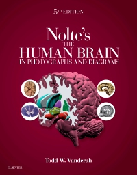
Nolte's The Human Brain in Photographs and Diagrams Elsevier eBook on VitalSource, 5th Edition
by Todd W. Vanderah, PhD
Elsevier eBook on VitalSource
ISBN:
9780323598194
Copyright:
2020
Publication Date:
12-21-2018
Page Count:
280
Imprint:
Elsevier
List Price:
$53.99
Most review copies are eBooks – how fast!
A review copy request is most likely to be fulfilled as an eBook on VitalSource rather than a print product, unless no eBook is available. eBooks become available in as little as a few hours. Print products will take between 7 and 10 days to arrive. To request a print copy, please contact us through the Evolve Support Center for further assistance or contact your Elsevier Sales Rep.
In the 5th Edition of this highly accessible atlas, Dr. Todd Vanderah continues the mission of his esteemed colleague, Dr. John "Jack" Nolte, to clearly depict and explain the challenging subject of neuroanatomy. Designed to promote a rapid understanding of complex concepts, Nolte's The Human Brain in Photographs and Diagrams, Fifth Edition combines easy-to-digest coverage of the brain, spinal cord, and brainstem with carefully selected visuals to cover all aspects of the information needed for success in coursework, on exams, and in clerkships and clinical practice.
-
- Features more than 600 high-quality figures including brain sections (transverse, coronal, axial, sagittal), 3-D reconstructions, MRIs and angiography, illustrated pathways that help you visualize anatomical structures and neuropathology
- Presents a systemic series of unlabelled whole brain sections next to corresponding sections with important structures outlined and labelled
- Includes a NEW chapter: An Introduction to Neuropathology, as well as NEW review questions online
- Helps you understand the connections between functional systems with detailed diagrams that incorporate actual brain and spinal cord sections
- Features clinical content throughout that shows how neuroanatomy applies to clinical practice
- Discusses every labelled structure in the highly illustrated glossary at the end of the book
- Shows major structures and major transitions in higher magnification for greater detail, and features bold index entries to indicate particularly clear illustrations of a given structure
- Enhanced eBook version included with purchase. Your enhanced eBook allows you to access all of the text, figures, and references – including 68 bonus dissection videos – from the book on a variety of devices
-
1. External Anatomy of the Brain
1.1. Introduction
1.2. Brain and Spinal Cord
1.3. Multiple Views of a Single Brain
1.4. Lateral and Superior Surfaces
1.5. Inferior Surface
1.6. Medial and Inferior Surfaces
1.7. Insula
1.8. Cerebellum
1.9. Brainstem
2. Transverse Sections of the Spinal Cord
2.1. Introduction
2.2. Guided Tour
2.3. Enlargements of Selected Levels
2.3.1. S4
2.3.2. L2
2.3.3. T10
2.3.4. C8
2.3.5. C3
3. Transverse Sections of the Brainstem
3.1. Introduction
3.2. Schematic Cross Sections
3.3. Blood Supply
3.4. Cranial Nerve Nuclei
3.5. Guided Tour
3.6. Enlargements of Selected Levels
3.6.1. Spinomedullary junction
3.6.2. Caudal medulla
3.6.3. Rostral medulla
3.6.4. Pontomedullary junction
3.6.5. Caudal pons
3.6.6. Midpons
3.6.7. Rostral pons
3.6.8. Caudal midbrain
3.6.9. Rostral midbrain
3.6.10. Far rostral midbrain
4. Building a Brain: Three-Dimensional Reconstructions
4.1. Introduction
4.2. Step-by-Step Reconstruction
5. Coronal Sections
5.1. Introduction
5.2. Guided Tour
5.3. Enlargements of Selected Levels
5.3.1. Striatum, anterior limb of the internal capsule
5.3.2. Anterior commissure
5.3.3. Anterior thalamus, ansa lenticularis
5.3.4. Amygdala, mammillary bodies
5.3.5. Anterior hippocampus, subthalamic nucleus
5.3.6. Posterior thalamus, red nucleus
5.3.7. Posterior thalamus, geniculate nuclei
5.3.8. Posterior thalamus, pulvinar
5.3.9. Atrium of the lateral ventricle
6. Axial Sections
6.1. Introduction
6.2. Guided Tour
6.3. Enlargements of Selected Levels
6.3.1. Uncus, optic chiasm
6.3.2. Hypothalamus, superior cerebellar peduncle
6.3.3. Striatum, deep cerebellar nuclei
6.3.4. Anterior commissure, subthalamic nucleus
6.3.5. Interventricular foramen, posterior commissure
6.3.6. Midthalamus
6.3.7. Roof of the third ventricle
6.3.8. Transverse fissure, internal cerebral veins
7. Sagittal Sections
7.1. Introduction
7.2. Guided Tour
7.3. Enlargements of Selected Levels
7.3.1. Putamen, hippocampus
7.3.2. Hippocampus, amygdala
7.3.3. Globus pallidus, internal capsule, thalamus
7.3.4. Caudate nucleus, dentate nucleus
7.3.5. Mammillothalamic tract, superior cerebellar peduncle
7.3.6. Medial thalamus, column of the fornix
7.3.7. Mammillary body, body of the fornix
7.3.8. Near the midline
8. Functional Systems
8.1. Detailed Contents
8.2. Long Tracts of the Spinal Cord and Brainstem
8.3. Sensory Systems of the Brainstem and Forebrain
8.4. Cranial Nerve Motor Nuclei
8.5. Visceral Afferents and Efferents
8.6. Basal Ganglia
8.7. Cerebellum
8.8. Thalamus and Cerebral Cortex
8.9. Hypothalamus and Limbic System
8.10. Chemically Coded Neuronal Systems
9. Clinical Imaging
9.1. Introduction
9.2. Computed Tomography
9.2.1. Without contrast
9.2.2. With contrast
9.2.3. Pathology
9.3. Magnetic Resonance Imaging (MRI)
9.3.1. Introduction
9.3.2. Coronal T1-weighted MRIs
9.3.3. Axial T1-weighted MRIs
9.3.4. Sagittal and parasaggital T1-weighted MRIs
9.3.5. Pathology
9.4. Angiography
9.4.1. Introduction
9.4.2. Internal carotid angiograms
9.4.3. Vertebral-basilar angiograms
9.4.4. Pathology
10. Neuropathology Imaging
Glossary -
-
Ways of Reading
- The appearance of the text and page layout can be modified according to the capabilities of the reading system (font family and font size, spaces between paragraphs, sentences, words, and letters, as well as color of background and text)
- This e-publication is accessible to the full extent that the file format and types of content allow, on a specific reading device, by default, without necessarily including any additions such as textual descriptions of images or enhanced navigation
- No information about nonvisual reading is available
-
Conformance
- No information is available
-
Navigation
- Table of contents to all chapters of the text via links
- Page list to go to pages from the print source version
-
Rich Content
- No information is available
-
Hazards
- No information is available
-
Product Content
- No information is available
-
Legal Considerations
- No information is available
-
Additional Accessibility Information
- Page breaks included from the original print source
- For readers with color vision deficiency, use of color (e.g., in diagrams, graphics and charts, in prompts, or on buttons inviting a response) is not the sole means of graphical distinction or of conveying information
- E-publication includes basic navigation (usually less detailed than TOC-based navigation)
- Where links, controls or buttons are included in the content, the purpose or functionality of each link, control or button is apparent from the associated text alone - or where it is unclear, separate link, control or button descriptions are provided
- All (or substantially all) textual matter is arranged in a single logical reading order (including text that is visually presented as separate from the main text flow, e.g., in boxouts, captions, tables, footnotes, endnotes, citations, etc.). Non-textual content is also linked from within this logical reading order. (Purely decorative non-text content can be ignored).
- The language of the text has been specified (e.g., via the HTML or XML lang attribute) to optimise text-to-speech (and other alternative renderings), both at the whole document level and, where appropriate, for individual words, phrases or passages in a different language.
-
Ways of Reading


Most review copies are eBooks – how fast!
A review copy request is most likely to be fulfilled as an eBook on VitalSource rather than a print product, unless no eBook is available. eBooks become available in as little as a few hours. Print products will take between 7 and 10 days to arrive. To request a print copy, please contact us through the Evolve Support Center for further assistance or contact your Elsevier Sales Rep.

 as described in our