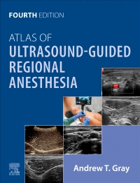

Atlas of Ultrasound-Guided Regional Anesthesia - Elsevier E-Book on VitalSource, 4th Edition
Elsevier eBook on VitalSource
Now $157.51
Written by one of the pioneers of the use of ultrasound to guide needle placement, Dr. Andrew T. Gray’s Atlas of Ultrasound-Guided Regional Anesthesia, 4th Edition, remains the reference of choice for clinicians who use ultrasound-guided technology to administer regional blocks. Step-by-step videos and images, board-style review questions, and discussions of new blocks make this highly respected title a must-have reference for anesthesiologists, pain medicine specialists, nurse anesthetists, and emergency physicians-anyone who requires expert guidance on the latest methods to ensure safe and effective needle placement.
-
- Helps ensure correct needle placement with numerous 3-D and long-axis views that clearly depict surrounding structures.
- Contains nine new chapters: Ultra High Frequency (UHF) Ultrasound; PENG Block; iPACK Block; External Oblique Intercoastal (EOI) Block; Serratus Anterior Plane Block; Erector Spinae Plane (ESP) Block; Suprazygomatic Maxillary Nerve Block; Ultrasound-Guided Percutaneous Cryoneurolysis for Acute Pain; Artificial Intelligence (AI) in Ultrasound-Guided Regional Anesthesia.
- Includes numerous high-quality anatomy illustrations, photographs demonstrating patient positioning, and ultrasound images demonstrating anatomy and needle placement.
- Features an expanded video collection with step-by-step guidance of ultrasound-guided techniques, including video flips for PENG, rectus sheath, maxillary nerve, sciatic nerve blocks, and more.
- Includes newly revised, board-style review questions to help test your knowledge and prepare you for the ABA exam.
-
SECTION 1 Introduction to Ultrasound Imaging
1. Ultrasound
2. Speed of Sound
3. Attenuation
4. Reflection
5. Beam Width (Slice Thickness)
6. Anisotropy
7. Spatial Compound Imaging
8. Doppler Imaging
9. Ultrasound Transducers
10. Transducer Manipulation
11. Needle Imaging
12. Approach and Techniques
13. Sonographic Signs of Successful Injections
14. Continuous Peripheral Nerve Blocks
15. Three-Dimensional Ultrasound
16. Ultra-High Frequency Ultrasound
SECTION 2 Structures
17. Anatomic Structures
18. Skin and Subcutaneous Tissue
19. Peripheral Nerves
20. Muscle
21. Tendons and Ligaments
22. Arteries
23. Veins
24. Bone
25. Pleura
26. Peritoneum
SECTION 3 Upper Extremity Blocks
27. Interscalene and Superior Trunk Blocks
28. Supraclavicular Block
29. Suprascapular Nerve Block (Anterior Approach, above the Clavicle)
30. Infraclavicular Block
31. Axillary Block
32. Musculocutaneous Nerve Block
33. Forearm Blocks
34. Radial Nerve Block
35. Median Nerve Block
36. Ulnar Nerve Block
SECTION 4 Lower Extremity Blocks
37. Lateral Femoral Cutaneous Nerve Block
38. Fascia Iliaca Block
39. Femoral Nerve Block
40. Adductor Canal Block
41. Obturator Nerve Block
42. Sciatic Nerve Block
43. Popliteal Block
44. Posterior Femoral Cutaneous Nerve Block
45. Pericapsular Nerve Group (PENG) Block for Hip Analgesia
46. Infiltration of Local Anesthetic between the Popliteal Artery and Capsule of Knee (iPACK) Block
47. Ankle Blocks
48. Saphenous Nerve Block in the Leg
49. Deep Peroneal Nerve Block
50. Superficial Peroneal Nerve Block
51. Sural Nerve Block
52. Tibial Nerve Block
SECTION 5 Trunk Blocks
53. Intercostal Nerve Block
54. External Oblique Intercostal Plane Block
55. Serratus Anterior Plane Block
56. Pectoral Nerve Block (The PECS Block)
57. Transversus Thoracis Muscular Plane Block (Parasternal Block)
58. Rectus Sheath Block
59. Ilioinguinal and Iliohypogastric Nerve Blocks
60. Transversus Abdominis Plane Block
61. Erector Spinae Plane Block
62. Thoracic Paravertebral Block
63. Neuraxial Block
64. Caudal Epidural Block
SECTION 6 Head and Neck Blocks
65. Supraorbital Nerve Block
66. Mental Nerve Block
67. Suprazygomatic Maxillary Nerve Block
68. Superior Laryngeal Nerve Block
69. Transtracheal Block
70. Greater Occipital Nerve Block
71. Cervical Plexus Block
72. Stellate Ganglion Block (Cervicothoracic Sympathetic Ganglion Block)
SECTION 7 Safety Issues and New Technology
73. Safety: Practical Techniques to Prevent Complications During Ultrasound-Guided Nerve Blocks
74. Ultrasound-Guided Percutaneous Cryoneurolysis for Acute Pain
75. Artificial Intelligence in Ultrasound-Guided Regional Anesthesia -
-
Ways of Reading
- The appearance of the text and page layout can be modified according to the capabilities of the reading system (font family and font size, spaces between paragraphs, sentences, words, and letters, as well as color of background and text)
- This e-publication is accessible to the full extent that the file format and types of content allow, on a specific reading device, by default, without necessarily including any additions such as textual descriptions of images or enhanced navigation
- No information about nonvisual reading is available
-
Conformance
- No information is available
-
Navigation
- Table of contents to all chapters of the text via links
- Page list to go to pages from the print source version
-
Rich Content
- No information is available
-
Hazards
- No information is available
-
Product Content
- No information is available
-
Legal Considerations
- No information is available
-
Additional Accessibility Information
- Content is enhanced with ARIA roles to optimize organization and facilitate navigation
- Page breaks included from the original print source
- For readers with color vision deficiency, use of color (e.g., in diagrams, graphics and charts, in prompts, or on buttons inviting a response) is not the sole means of graphical distinction or of conveying information
- E-publication includes basic navigation (usually less detailed than TOC-based navigation)
- Where links, controls or buttons are included in the content, the purpose or functionality of each link, control or button is apparent from the associated text alone - or where it is unclear, separate link, control or button descriptions are provided
- All (or substantially all) textual matter is arranged in a single logical reading order (including text that is visually presented as separate from the main text flow, e.g., in boxouts, captions, tables, footnotes, endnotes, citations, etc.). Non-textual content is also linked from within this logical reading order. (Purely decorative non-text content can be ignored).
- The language of the text has been specified (e.g., via the HTML or XML lang attribute) to optimise text-to-speech (and other alternative renderings), both at the whole document level and, where appropriate, for individual words, phrases or passages in a different language.
-
Ways of Reading


 as described in our