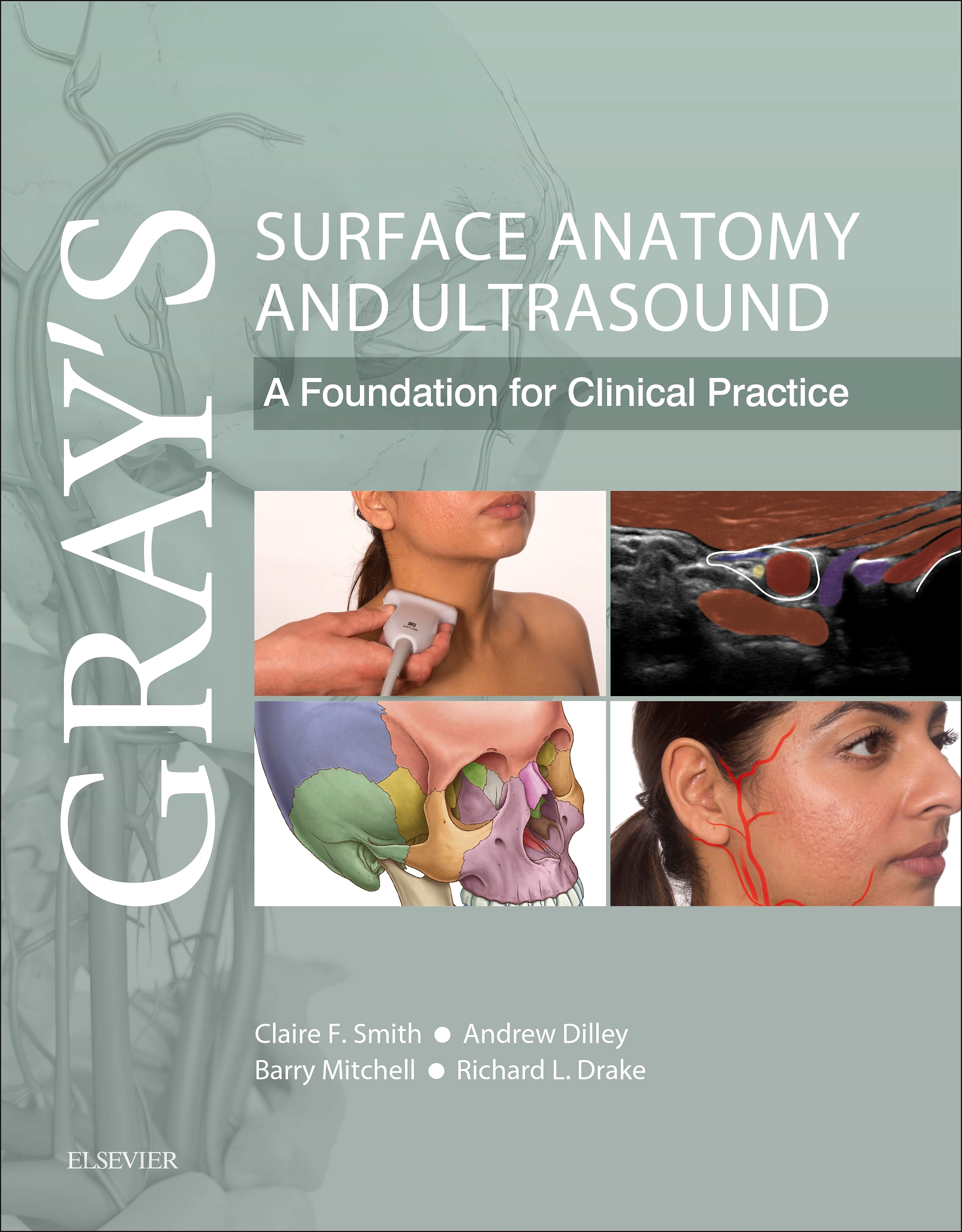
Gray’s Surface Anatomy and Ultrasound, 1st Edition
Paperback

Now $44.63
A concise, superbly illustrated (print + electronic) textbook that brings together a reliable, clear and up to date guide to surface anatomy and its underlying gross anatomy, combined with a practical application of ultrasound and other imaging modalities.
A thorough understanding of surface anatomy remains a critical part of clinical practice, but with improved imaging technology, portable ultrasound is also fast becoming integral to routine clinical examination and effective diagnosis.
This unique new text combines these two essential approaches to effectively understanding clinical anatomy and reflects latest approaches within modern medical curricula. It is tailored specifically to the needs of medical students and doctors in training and will also prove invaluable to the wide range of allied health students and professionals who need a clear understanding of visible and palpable anatomy combined with anatomy as seen on ultrasound.
- Concise text and high quality illustrations, photographs, CT, MRI and ultrasound scans provide a clear, integrated understanding of the anatomical basis for modern clinical practice
- Highly accessible and at a level appropriate for medical students and a wide range of allied health students and professionals
- Reflects current curriculum trend of heavily utilizing living anatomy and ultrasound to learn anatomy
- Supplementary video content showing live ultrasound scans and guided areas of surface anatomy to bring content to life and reflect current teaching and clinical settings
- An international advisory panel appointed to add expertise and ensure relevance to the variety of medical and allied health markets
- Inclusion of latest ultrasound image modalities
- Designed to complement and enhance the highly successful Gray’s family of texts/atlases although also effective as a stand-alone or alongside other established anatomy resources
-
1. Introduction
Conceptual overview
Surface anatomy
Anatomical position and planes
Anatomical terms
Movement
Fascia
Skin
Skin color
Dermatomes and myotomes
Natural variation
Palpation and percussion
Ultrasound
Ultrasound theory
Doppler
Types of transducer
Imaging planes
Screen orientation
Ergonomics
Manipulating the transducer
Short-axis and long-axis views
Image terminology
Appearance of tissues
2. Thorax
Conceptual overview
Surface anatomy
Bones
Muscles
Breast
Thoracic cavity
Ultrasound
Anterior muscles of the thorax and lungs
Heart
Video 2.1 Colour Doppler ultrasound image sequence of the heart – apical view.
3. Abdomen
Conceptual overview
Surface anatomy
Bones
Abdominal regions
Muscles
Viscera
Ultrasound
Anterior abdominal musculature
Gastrointestinal tract
Liver
Kidney
Spleen
Pancreas
Vasculature
Video 3.1 B-mode ultrasound image sequence of the jejunum – transverse view.
4. Pelvis and perineum
Conceptual overview
Surface anatomy
Bones
Muscles
Viscera
Perineum
Pregnancy
Ultrasound
Male pelvis
Female pelvis
Video 4.1 Colour Doppler ultrasound image sequence of the bladder – mid-sagittal view.
5. Back
Conceptual overview
Surface anatomy
Curvatures
Bones
Ligaments
Joints
Muscles
Movements
Vertebral canal and spinal nerves
Ultrasound
6. Upper limb
Conceptual overview
Surface anatomy
Shoulder
Axilla
Arm
Forearm
Hand
Neurovascular structures
Ultrasound
Scalene triangle
Shoulder region
Deltoid muscle
Rotator cuff muscles
Anterior arm
Posterior arm
Elbow
Anterior forearm
Posterior forearm
Hand
Video 6.1 B-mode ultrasound image sequence of the long flexor tendons immediately proximal to the wrist – long-axis view.
7. Lower limb
Conceptual overview
Surface anatomy
Gluteal region
Thigh
Knee joint
Leg
Foot
Neurovascular structures
Ultrasound
Gluteal region
Femoral triangle
Anterior thigh
Knee
Medial thigh and adductor canal
Posterior thigh and popliteal fossa
Anterior leg
Posterior leg
Lateral leg
Video 7.1 Colour Doppler ultrasound image sequence of the femoral artery and vein in the thigh – long-axis view.
8. Head and neck
Conceptual overview
Surface anatomy
Head
Neck
Lymph
Neurovascular
Ultrasound
Eye
Parotid gland
Submandibular gland
Floor of the oral cavity
Carotid system
Thyroid gland
Posterior triangle of neck
Video 8.1 Colour Doppler ultrasound image sequence of the internal and external carotid arteries (red) and internal jugular vein (blue) in the neck – short-axis view.


 as described in our