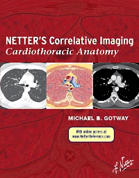
Netter’s Correlative Imaging: Cardiothoracic Anatomy, 1st Edition
Hardcover

Now $195.29
Cardiothoracic Anatomy, the third title in the brand-new Netter’s Correlative Imaging series, provides exceptional visual guidance for thoracic, chest wall, lung, and heart anatomy. Dr. Michael Gotway presents Netter’s beautiful and instructive paintings and illustrated cross sections created in the Netter style side-by-side with high-quality patient images from breath-hold cardiac MR, multislice thoracic CT, and CT coronary angiography to help you visualize the anatomy section by section. With in-depth coverage and concise descriptive text for at-a-glance information and access to correlated images online, this atlas is a comprehensive reference that’s ideal for today’s busy imaging specialists.
-
- View thoracic, chest wall, lung, and heart anatomy in breath-hold cardiac MR, multislice thoracic CT, and CT coronary angiography, each image complemented by a detailed illustration in the instructional and aesthetic Netter style.
- Find anatomical landmarks quickly and easily through comprehensive labeling and concise text highlighting key points related to the illustration and image pairings.
-
Part 1 Thoracic Anatomy
Chapter 1 Overview of Thoracic Anatomy
Chapter 2 Thoracic Soft Tissue and Lung
Axial
Coronal
Chapter 3 Pulmonary Anatomy and Variants
Axial
Coronal
Chapter 4 Thoracic Lymph Nodes
Axial
Chapter 5 Cisterna Chyli and Thoracic Duct
Axial
Coronal
Chapter 6 Venous Anatomy and Variants
Axial
Coronal
Sagittal
Part 2 Cardiac Anatomy
Chapter 7 Overview of Cardiac Anatomy
Chapter 8 Cardiac Anatomy Using CT
Axial
2-chamber
3-chamber
4-chamber
Short Axis
Chapter 9 Cardiac Anatomy Using MR
Axial
2-chamber
3-chamber
4-chamber
Short Axis

 as described in our