ISBN:
9780323312004
Copyright:
2016
Publication Date:
02-19-2015
Imprint:
Mosby
List Price:
$282.99
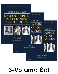
Merrill's Atlas of Radiographic Positioning and Procedures - Elsevier eBook on VitalSource, 13th Edition
by Bruce W. Long, MS, RT(R)(CV), FASRT, FAEIRS, Jeannean Hall Rollins, MRC, BSRT(R)(CV)(M) and Barbara J. Smith, MS, RT(R)(QM), FASRT, FAEIRS
Elsevier eBook on VitalSource
ISBN:
9780323312004
Copyright:
2016
Publication Date:
02-19-2015
Imprint:
Mosby
List Price:
$282.99
Most review copies are eBooks – how fast!
A review copy request is most likely to be fulfilled as an eBook on VitalSource rather than a print product, unless no eBook is available. eBooks become available in as little as a few hours. Print products will take between 7 and 10 days to arrive. To request a print copy, please contact us through the Evolve Support Center for further assistance or contact your Elsevier Sales Rep.
More than 400 projections make it easier to learn anatomy, properly position the patient, set exposures, and take high-quality radiographs! With Merrill's Atlas of Radiographic Positioning & Procedures, 13th Edition, your students will develop the skills to produce clear radiographic images to help physicians make accurate diagnoses. It separates anatomy and positioning information by bone groups or organ systems — using full-color illustrations to show anatomical anatomy, and CT scans and MRI images to help in learning cross-section anatomy. Merrill's Atlas is not just the gold standard in radiographic positioning texts, and the most widely used, but also an excellent review in preparing for ARRT and certification exams!
-
- Includes a unique new section on working with and positioning obese patients.
- Offers coverage of one new compensating filter.
- Provides collimation sizes and other key information for each relevant projection.
- Features more CT and MRI images to enhance your understanding of cross-sectional anatomy and prepare you for the Registry exam.
- Offers additional digital images in each chapter, including "stitching" for long-length images of the spine and lower limb.
- Standardized image receptor sizes use English measurements with metric in parentheses.
- Depicts the newest equipment with updated photographs and images.
-
- UNIQUE! Collimation sizes and other key information are provided for each relevant projection.
- NEW! Coverage of the latest advances in digital imaging also includes more digital radiographs with greater contrast resolution of pertinent anatomy.
- NEW positioning photos show current digital imaging equipment and technology.
- Comprehensive, full-color coverage of anatomy and positioning makes Merrill's Atlas the most in-depth text and reference available for radiography students and practitioners.
- Coverage of common and unique positioning procedures includes special chapters on trauma, surgical radiography, geriatrics/pediatrics, and bone densitometry, to help prepare students for the full scope of situations they will encounter.
- Numerous CT and MRI images enhance comprehension of cross-sectional anatomy and help in preparing for the Registry examination.
- UPDATED! Combined coverage of skull, facial bones, and paranasal sinuses enhances learning of the anatomy of these structures, and the Summary of Pathology table now includes facial bone fractures and sinus pathologies.
- UPDATED Mammography chapter reflects the evolution to digital mammography, as well as innovations in breast biopsy procedures.
- UPDATED Pediatric Imaging chapter addresses care for the patient with autism, strategies for visit preparation, appropriate communication, and environmental considerations.
- UPDATED Geriatric Radiography chapter describes how to care for the patient with Alzheimer’s Disease and other related conditions.
- Expanded Summary of Pathology table now includes common male reproductive system pathologies.
- UPDATED coverage addresses contrast arthrography procedures, trauma radiography practices, plus current patient preparation, contrast media used, and the influence of digital technologies.
- Summary tables provide quick access to projection overviews, guides to anatomy, pathology tables for bone groups and body systems, and exposure technique charts.
- Bulleted lists provide clear instructions on how to correctly position the patient and body part when performing procedures.
-
VOLUME 1
1. Preliminary Steps in Radiography
2. Compensating Filters
3. General Anatomy and Radiographic Positioning Terminology
4. Upper Limb
5. Shoulder Girdle
6. Lower Limb
7. Pelvis and Upper Femora
8. Vertebral Column
9. Bony Thorax
10. Thoracic Viscera
VOLUME 2
11. Long Bone Measurement
12. Contrast Arthrography
13. Trauma Radiography
14. Mouth and Salivary Glands
15. Anterior Part of Neck
16. Abdomen
17. Digestive System: Alimentary Canal
18. Urinary System and Venipuncture
19. Reproductive System
20. Skull, Facial Bones, and Paranasal Sinuses
21. Mammography
VOLUME 3
22. Central Nervous System
23. Vascular, Cardiac, and Interventional Radiography
24. Pediatric Imaging
25. Geriatric Radiography
26. Mobile Radiography
27. Surgical Radiography
28. Sectional Anatomy for Radiographers
29. Computed Tomography
30. Magnetic Resonance Imaging
31. Diagnostic Medical Sonography
32. Nuclear Medicine
33. Bone Densitometry
34. Radiation Oncology -
No accessibility information is available for this product.
Most review copies are eBooks – how fast!
A review copy request is most likely to be fulfilled as an eBook on VitalSource rather than a print product, unless no eBook is available. eBooks become available in as little as a few hours. Print products will take between 7 and 10 days to arrive. To request a print copy, please contact us through the Evolve Support Center for further assistance or contact your Elsevier Sales Rep.
 as described in our