ISBN:
9780323881777
Copyright:
2025
Publication Date:
05-07-2024
Page Count:
270
Imprint:
Elsevier
List Price:
$90.99

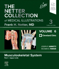
The Netter Collection of Medical Illustrations: Musculoskeletal System, Volume 6, Part I - Upper Limb - Elsevier E-Book on VitalSource, 3rd Edition
by Joseph Iannotti, MD, PhD, Richard Parker, MD, Tom Mroz, MD, Brendan Patterson, MD and Abby Abelson, MD
Elsevier eBook on VitalSource
ISBN:
9780323881777
Copyright:
2025
Publication Date:
05-07-2024
Page Count:
270
Imprint:
Elsevier
List Price:
$90.99
Most review copies are eBooks – how fast!
A review copy request is most likely to be fulfilled as an eBook on VitalSource rather than a print product, unless no eBook is available. eBooks become available in as little as a few hours. Print products will take between 7 and 10 days to arrive. To request a print copy, please contact us through the Evolve Support Center for further assistance or contact your Elsevier Sales Rep.
Offering a concise, highly visual approach to the basic science and clinical pathology of the musculoskeletal system, this updated volume in The Netter Collection of Medical Illustrations (the CIBA "Green Books") contains unparalleled didactic illustrations reflecting the latest medical knowledge. Revised by Drs. Joseph Iannotti, Richard Parker, and other experts from the Cleveland Clinic, The Upper Limb, Part 1 of Musculoskeletal System, Volume 6, integrates core concepts of anatomy, physiology, and other basic sciences with common clinical correlates across health, medical, and surgical disciplines. Classic Netter art, updated and new illustrations, and modern imaging continue to bring medical concepts to life and make this timeless work an essential resource for students, clinicians, and educators.
-
- Provides a highly visual guide to the shoulder, upper arm and elbow, forearm and wrist, and hand and finger, from basic science and anatomy to orthopaedics and rheumatology
- Covers new topics including surgical management of irreparable tears: supraspinatus and infraspinatus cuff, and subscapularis
- Provides a concise overview of complex information by seamlessly integrating anatomical and physiological concepts using practical clinical scenarios
- Shares the experience and knowledge of Drs. Joseph P. Iannotti, Richard D. Parker, and esteemed colleagues from the Cleveland Clinic, who clarify and expand on the illustrated concepts
- Compiles Dr. Frank H. Netter’s master medical artistry—an aesthetic tribute and source of inspiration for medical professionals for over half a century—along with new art in the Netter tradition for each of the major body systems, making this volume a powerful and memorable tool for building foundational knowledge and educating patients or staff
- NEW! An eBook version is included with purchase. The eBook allows you to access all of the text, figures, and references, with the ability to search, make notes and highlights, and have content read aloud
-
SECTION 1 SHOULDER
Anatomy
1.1 Scapula and Humerus: Posterior View
1.2 Scapula and Humerus: Anterior View
1.3 Clavicle
1.4 Ligaments
1.5 Glenohumeral Arthroscopic Anatomy
1.6 Glenohumeral Arthroscopic Anatomy (Continued)
1.7 Anterior Muscles
1.8 Anterior Muscles: Cross Section
1.9 Posterior Muscles
1.10 Posterior Muscles: Cross Section
1.11 Muscles of Rotator Cuff
1.12 Muscles of Rotator Cuff: Cross Sections
1.13 Axilla Dissection: Anterior View
1.14 Axilla: Posterior Wall and Cord
1.15 Deep Neurovascular Structures and Intervals
1.16 Axillary and Brachial Arteries
1.17 Axillary Artery and Anastomoses Around Scapula
1.18 Brachial Plexus
1.19 Peripheral Nerves: Dermatomes
1.20 Peripheral Nerves: Sensory Distribution and Neuropathy in Shoulder
Clinical Problems and Correlations
Fractures and Dislocation
1.21 Proximal Humeral Fractures: Neer Classification
1.22 Proximal Humeral Fractures: Two-Part Tuberosity Fracture
1.23 Proximal Humeral Fractures: Two Part Surgical Neck Fracture and Humeral Head Dislocation
1.24 Proximal Humeral Fractures: Valgus-Impacted Four-Part Fracture
1.25 Proximal Humeral Fractures: Displaced Four-Part Fractures with Articular Head Fracture
1.26 Proximal Humeral Fractures: Reverse Total Shoulder Replacement
1.27 Anterior Dislocation of Glenohumeral Joint: Anterior Dislocation Types and Stimson Maneuver
1.28 Anterior Dislocation of Glenohumeral Joint: Pathologic Lesions
1.29 Anterior Dislocation of Glenohumeral Joint: Imaging
1.30 Posterior Dislocation of Glenohumeral Joint
1.31 Posterior Dislocation of Glenohumeral Joint (Continued)
1.32 Acromioclavicular and Sternoclavicular Dislocation
1.33 Fractures of the Clavicle
1.34 Fractures of the Clavicle and Scapula
Common Soft Tissue Disorders
1.35 Calcific Tendonitis
1.36 Frozen Shoulder: Clinical Presentation
1.37 Frozen Shoulder: Risk Factors and Diagnostic Tests
1.38 Biceps, Tendon Tears, and SLAP Lesions: Presentation and Physical Examination
1.39 Biceps, Tendon Tears, and SLAP Lesions: Types of Tears
1.40 Acromioclavicular Joint Arthritis
1.41 Impingement Syndrome and the Rotator Cuff: Presentation and Diagnosis
1.42 Impingement Syndrome and the Rotator Cuff: Radiologic and Arthroscopic Imaging
1.43 Rotator Cuff Tears: Physical Examination
1.44 Supraspinatus and Infraspinatus Rotator Cuff Tears: Imaging
1.45 Supraspinatus and Infraspinatus Rotator Cuff Tears: Surgical Management
1.46 Supraspinatus and Infraspinatus Cuff Tear: Surgical Management Superior Capsular Reconstruction and Balloon Arthroplasty
1.47 Supraspinatus and Infraspinatus Cuff Tear: Surgical Management Latissimus and Trapezius Transfers
1.48 Management of Subscapularis Rotator Cuff Tears
1.49 Management of Irreparable Subscapularis Tears: Pectoralis and Latissimus Transfers
1.50 Osteoarthritis of the Glenohumeral Joint
1.51 Osteoarthritis of the Glenohumeral Joint: Surgical Imaging
1.52 Avascular Necrosis of the Humeral Head
1.53 Rheumatoid Arthritis of the Glenohumeral Joint: Radiographic Presentations and Treatment Options
1.54 Rheumatoid Arthritis of the Glenohumeral Joint: Conservative Humeral Head Surface Replacement
1.55 Rotator Cuff–Deficient Arthritis (Rotator Cuff Tear Arthropathy): Physical Findings and Appearance
1.56 Rotator Cuff–Deficient Arthritis (Rotator Cuff Tear Arthropathy): Radiographic Findings
1.57 Rotator Cuff–Deficient Arthritis (Rotator Cuff Tear Arthropathy): Radiographic Findings (Continued)
1.58 Neurologic Conditions of the Shoulder: Suprascapular Nerve
1.59 Neurologic Conditions of the Shoulder: Long Thoracic and Spinal Accessory Nerves
Amputation
1.60 Amputation of Upper Arm and Shoulder, 61
Injections, Basic Rehabilitation, and Surgical Approaches
1.61 Shoulder Injections
1.62 Basic, Passive, and Active-Assisted Range of Motion Exercises
1.63 Basic Shoulder-Strengthening Exercises
1.64 Basic Shoulder Strengthening Exercises (Continued)
1.65 Common Surgical Approaches to the Shoulder
SECTION 2 UPPER ARM AND ELBOW
Anatomy
2.1 Topographic Anatomy
2.2 Anterior and Posterior Views of Humerus
2.3 Elbow Joint: Bones
2.4 Elbow Joint: Radiographs
2.5 Elbow Ligaments: Anterior Views
2.6 Elbow Ligaments: Lateral and Medial Views
2.7 Muscles Origins and Insertions
2.8 Muscles: Anterior Views
2.9 Muscles: Posterior Views
2.10 Cross Sectional Anatomy of Upper Arm
2.11 Cross Sectional Anatomy of Elbow
2.12 Cutaneous Nerves and Superficial Veins
2.13 Cutaneous Innervation
2.14 Musculocutaneous Nerve of Upper Arm and Elbow
2.15 Radial Nerve
2.16 Brachial Artery In Situ
2.17 Brachial Artery and Anastomoses around Elbow
Clinical Problems and Correlations
2.18 Physical Examination and Range of Motion
Fractures and Dislocation
2.19 Humeral Shaft Fractures
2.20 Injury to the Elbow
2.21 Fracture of Distal Humerus
2.22 Fracture of Distal Humerus: Total Elbow Arthroplasty
2.23 Fracture of Distal Humerus: Capitellum
2.24 Fracture of Head and Neck of Radius
2.25 Fracture of Head and Neck of Radius: Imaging
2.26 Fracture of Olecranon
2.27 Dislocation of Elbow Joint
2.28 Dislocation of Elbow Joint (Continued)
2.29 Injuries in Children: Supracondylar Humerus Fractures
2.30 Injuries in Children: Elbow
2.31 Injuries in Children: Subluxation of Radial Head
2.32 Complications of Fracture
Common Soft Tissue Disorders
2.33 Arthritis: Open and Arthroscopic Elbow Debridement
2.34 Arthritis: Elbow Arthroplasty Options
2.35 Arthritis: Imaging of Total Elbow Arthroplasty Designs
2.36 Cubital Tunnel Syndrome: Sites of Compression
2.37 Cubital Tunnel Syndrome: Clinical Signs and Treatment
2.38 Epicondylitis and Olecranon Bursitis
2.39 Rupture of Biceps and Triceps Tendon
2.40 Medial Elbow and Posterolateral Rotatory Instability Tests
2.41 Osteochondritis Dissecans of the Elbow
2.42 Osteochondrosis of the Elbow (Panner Disease)
2.43 Congenital Dislocation of Radial Head
2.44 Congenital Radioulnar Synostosis
Injections, Basic Rehabilitation, and Surgical Approaches
2.45 Common Elbow Injections and Basic Rehabilitation
2.46 Surgical Approaches to the Upper Arm and Elbow
2.47 Surgical Approaches to the Upper Arm and Elbow (Continued)
SECTION 3 FOREARM AND WRIST
Anatomy
3.1 Topographic Anatomy
3.2 Bones of Forearm
3.3 Bones of Wrist
3.4 Radiologic Findings of Wrist
3.5 Ligaments of Wrist
3.6 Arthroscopy of Wrist
3.7 Muscles of Forearm (Superficial Layer): Anterior View
3.8 Muscles of Forearm (Intermediate and Deep Layers): Anterior View
3.9 Muscles of Forearm (Superficial and Deep Layers): Posterior View
3.10 Cross-Sectional Anatomy of Right Forearm
3.11 Cross-Sectional Anatomy of Wrist
3.12 Muscles of Forearm: Origins and Insertions
3.13 Blood Supply of Forearm
3.14 Median Nerve of Forearm
3.15 Ulnar Nerve of Forearm
3.16 Cutaneous Nerves of Forearm
Clinical Problems and Correlations
3.17 Carpal Tunnel Syndrome
3.18 Cubital Tunnel Syndrome
3.19 Fracture of Distal Radius: Colles Fracture
3.20 Fracture of Distal Radius: Radiology
3.21 Fracture of Distal Radius: Closed Reduction and Plaster Cast Immobilization of Colles Fracture
3.22 Fracture of Distal Radius: Radiology of Open Reduction and Internal Fixation
3.23 Fracture of Scaphoid: Presentation and Classification
3.24 Fracture of Scaphoid: Blood Supply and Treatment
3.25 Fracture of Scaphoid: Radiology
3.26 Fracture of Hamulus of Hamate
3.27 Dislocation of Carpus: Presentation and Treatment
3.28 Dislocation of Carpus: Radiology
3.29 Fracture of Both Forearm Bones
3.30 Fracture of Shaft of Ulna
3.31 Fracture of Shaft of Radius
3.32 Ganglion of Wrist
3.33 De Quervain Disease
3.34 Rheumatoid Arthritis of Wrist
3.35 Arthritis of Wrist
3.36 Kienböck Disease
3.37 Radial Longitudinal Deficiency: Forearm Manifestations
3.38 Radial Longitudinal Deficiency: Type II Hypoplastic Thumb
SECTION 4 HAND AND FINGER
Anatomy
4.1 Topographic Anatomy: Anterior View
4.2 Topographic Anatomy: Posterior View
4.3 Metacarpophalangeal and Interphalangeal Ligaments
4.4 Definitions of Hand Motion
4.5 Flexor and Extensor Tendons in Fingers
4.6 Flexor and Extensor Zones and Lumbrical Muscles
4.7 Wrist and Hand: Deep Dorsal Dissection
4.8 Wrist and Hand: Intrinsic Muscles
4.9 Spaces, Bursae, and Tendon and Lumbrical Sheaths of Hand
4.10 Wrist and Hand: Palmar Dissections
4.11 Vascular Supply of the Hand and Finger
4.12 Ulnar Nerve of Hand
4.13 Median Nerve of Hand
4.14 Radial Nerve of Hand
4.15 Skin and Subcutaneous Fascia: Anterior View
4.16 Skin and Subcutaneous Fascia: Posterior View
4.17 Lymphatic Drainage
4.18 Digits: Sectional Anatomy
4.19 Thumb: Sectional Anatomy
Degenerative and Systemic Disorders
4.20 Hand Involvement in Osteoarthritis
4.21 Hand Involvement in Rheumatoid Arthritis and Psoriatic Arthritis
4.22 Hand Involvement in Gouty Arthritis and Reiter Syndrome
4.23 Deformities of Thumb Joints: Metacarpophalangeal Deformities
4.24 Deformities of Thumb Joints: Carpometacarpal Osteoarthritis
4.25 Deformities of Thumb Joints: Ligament Replacement and Tendon Interposition Arthroplasty
4.26 Deformities of the Metacarpophalangeal Joints: Implant Resection Arthroplasty
4.27 Deformities of the Metacarpophalangeal Joints: Implant Resection Arthroplasty (Continued)
4.28 Deformities of the Metacarpophalangeal Joints: Implant Resection Arthroplasty (Continued)
4.29 Deformities of the Metacarpophalangeal Joints: Modular versus Implant Resection Arthroplasty
4.30 Deformities of Interphalangeal Joint: Radiographic Findings
4.31 Deformities of Interphalangeal Joint: Swan Neck and Boutonniere
4.32 Deformities of Interphalangeal Joint: Implant Resection Arthroplasty
4.33 Deformities of Interphalangeal Joint: Modular Versus Implant Resection Arthroplasty
4.34 Dupuytren Contracture: Presentation and Treatment
4.35 Dupuytren Contracture: Surgical Approach to Finger
Infections and Tendon Disorders
4.36 Cellulitis and Abscess
4.37 Tenosynovitis and Infection of Fascial Space
4.38 Tenosynovitis and Infection of Fascial Space (Continued)
4.39 Infected Wounds
4.40 Infection of Deep Compartments of Hand
4.41 Lymphangitis
4.42 Bier Block Anesthesia
4.43 Thumb Carpometacarpal Injection, Digital Block, and Flexor Sheath Injection
4.44 Tendon Disorders: Trigger Finger and Jersey Finger
4.45 Tendon Disorders: Repair of Tendon
Fractures and Dislocations
4.46 Fracture of Metacarpal Neck and Shaft
4.47 Fracture of Thumb Metacarpal Base
4.48 Fracture of Proximal and Middle Phalanges
4.49 Management of Fracture of Proximal and Middle Phalanges
4.50 Special Problems in Fracture of Middle and Proximal Phalanges
4.51 Thumb Ligament Injury and Dislocation
4.52 Carpometacarpal and Metacarpophalangeal Joint Injury
4.53 Dorsal and Palmar Interphalangeal Joint Dislocations
4.54 Treatment of Dorsal Interphalangeal Joint Dislocation
4.55 Injuries to the Fingertip
4.56 Rehabilitation after Injury to Hand and Fingers
Amputation and Replantation
4.57 Amputation of Phalanx
4.58 Amputation of Thumb and Deepening of Thenar Web Cleft
4.59 Thumb Lengthening Post Amputation
4.60 Microsurgical Instrumentation for Replantation
4.61 Debridement, Incisions, and Repair of Bone in Replantation of Digit
4.62 Repair of Blood Vessels and Nerves
4.63 Postoperative Dressing and Monitoring of Blood Flow
4.64 Replantation of Avulsed Thumb and Midpalm
4.65 Lateral Arm Flap for Defect of Thumb Web
4.66 Transfer of Great Toe to Thumb Site
Selected References -
-
Ways of Reading
- The appearance of the text and page layout can be modified according to the capabilities of the reading system (font family and font size, spaces between paragraphs, sentences, words, and letters, as well as color of background and text)
- This e-publication is accessible to the full extent that the file format and types of content allow, on a specific reading device, by default, without necessarily including any additions such as textual descriptions of images or enhanced navigation
- No information about nonvisual reading is available
-
Conformance
- No information is available
-
Navigation
- Table of contents to all chapters of the text via links
- Page list to go to pages from the print source version
-
Rich Content
- No information is available
-
Hazards
- No information is available
-
Product Content
- No information is available
-
Legal Considerations
- No information is available
-
Additional Accessibility Information
- Content is enhanced with ARIA roles to optimize organization and facilitate navigation
- Page breaks included from the original print source
- For readers with color vision deficiency, use of color (e.g., in diagrams, graphics and charts, in prompts, or on buttons inviting a response) is not the sole means of graphical distinction or of conveying information
- E-publication includes basic navigation (usually less detailed than TOC-based navigation)
- Where links, controls or buttons are included in the content, the purpose or functionality of each link, control or button is apparent from the associated text alone - or where it is unclear, separate link, control or button descriptions are provided
- All (or substantially all) textual matter is arranged in a single logical reading order (including text that is visually presented as separate from the main text flow, e.g., in boxouts, captions, tables, footnotes, endnotes, citations, etc.). Non-textual content is also linked from within this logical reading order. (Purely decorative non-text content can be ignored).
- The language of the text has been specified (e.g., via the HTML or XML lang attribute) to optimise text-to-speech (and other alternative renderings), both at the whole document level and, where appropriate, for individual words, phrases or passages in a different language.
-
Ways of Reading

Most review copies are eBooks – how fast!
A review copy request is most likely to be fulfilled as an eBook on VitalSource rather than a print product, unless no eBook is available. eBooks become available in as little as a few hours. Print products will take between 7 and 10 days to arrive. To request a print copy, please contact us through the Evolve Support Center for further assistance or contact your Elsevier Sales Rep.

 as described in our