ISBN:
9780323930970
Copyright:
2025
Publication Date:
03-01-2024
Page Count:
330
Imprint:
Elsevier
List Price:
$101.99

The Netter Collection of Medical Illustrations: Cardiovascular System, Volume 8 - Elsevier E-Book on VitalSource, 3rd Edition
by Jamie B. Conti, MD and C. Richard Conti, MD
Elsevier eBook on VitalSource
ISBN:
9780323930970
Copyright:
2025
Publication Date:
03-01-2024
Page Count:
330
Imprint:
Elsevier
List Price:
$101.99
Most review copies are eBooks – how fast!
A review copy request is most likely to be fulfilled as an eBook on VitalSource rather than a print product, unless no eBook is available. eBooks become available in as little as a few hours. Print products will take between 7 and 10 days to arrive. To request a print copy, please contact us through the Evolve Support Center for further assistance or contact your Elsevier Sales Rep.
**Selected for 2026 Doody's Core Titles in Cardiology**
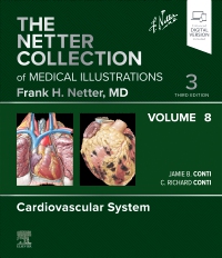
Offering a concise, highly visual approach to the basic science and clinical pathology of the cardiovascular system, this updated volume in The Netter Collection of Medical Illustrations (the CIBA "Green Books") contains unparalleled didactic illustrations reflecting the latest medical knowledge. Revised by Drs. Jamie B. Conti and C. Richard Conti, Cardiovascular System, Volume 8 integrates core concepts of anatomy, physiology, and other basic sciences with common clinical correlates across health, medical, and surgical disciplines. Classic Netter art, updated and new illustrations, and modern imaging continue to bring medical concepts to life and make this timeless work an essential resource for students, clinicians, and educators.
Offering a concise, highly visual approach to the basic science and clinical pathology of the cardiovascular system, this updated volume in The Netter Collection of Medical Illustrations (the CIBA "Green Books") contains unparalleled didactic illustrations reflecting the latest medical knowledge. Revised by Drs. Jamie B. Conti and C. Richard Conti, Cardiovascular System, Volume 8 integrates core concepts of anatomy, physiology, and other basic sciences with common clinical correlates across health, medical, and surgical disciplines. Classic Netter art, updated and new illustrations, and modern imaging continue to bring medical concepts to life and make this timeless work an essential resource for students, clinicians, and educators.
-
- Provides a highly visual guide to the heart and blood vessels, from basic science, anatomy, and physiology to pathology and injury
- Covers new diagnostics and therapeutics, including timely topics like intracardiac echocardiography, optical coherence tomography, radiation dose concerns, and coronary artery spasm
- Provides a concise overview of complex information by integrating anatomical and physiological concepts with clinical scenarios
- Compiles Dr. Frank H. Netter’s master medical artistry—an aesthetic tribute and source of inspiration for medical professionals for over half a century—along with new art in the Netter tradition for each of the major body systems, making this volume a powerful and memorable tool for building foundational knowledge and educating patients or staff
- NEW! An eBook version is included with purchase. The eBook allows you to access all of the text, figures, and references, with the ability to search, make notes and highlights, and have content read aloud
-
SECTION 1 ANATOMY
1.1 Thorax: Lungs in Situ
1.2 Thorax: Heart in Situ
1.3 Thorax: Mediastinum
1.4 Thorax: Pericardial Sac
1.5 Exposure of the Heart: Anterior Exposure
1.6 Exposure of the Heart: Base and Diaphragmatic Surfaces
1.7 Atria and Ventricles: Right Atrium and Right Ventricle
1.8 Atria and Ventricles: Left Atrium and Left Ventricle
1.9 Atria and Ventricles: Atria, Ventricles, and Interventricular Septum
1.10 Valves: Cardiac Valves Open and Closed
1.11 Valves: Valves and Fibrous Skeleton of Heart
1.12 Specialized Conduction System of Heart
1.13 Coronary Arteries and Cardiac Veins: Sternocostal and Diaphragmatic Surfaces
1.14 Coronary Arteries and Cardiac Veins: Arteriovenous Variations
1.15 Innervation of Heart: Nerves of Heart
1.16 Innervation of Heart: Schema of Innervation
SECTION 2 PHYSIOLOGY
2.1 Cardiovascular Examination: Cardiac Cycle
2.2 Cardiovascular Examination: Important Components
2.3 Cardiovascular Examination: Positions for Cardiac Auscultation
2.4 Cardiovascular Examination: Areas of Cardiac Auscultation
2.5 Cardiovascular Examination: Murmurs
2.6 Neural and Humoral Regulation of Cardiac Function
2.7 Physiologic Changes During Pregnancy
2.8 Cardiac Catheterization: Vascular Access
2.9 Cardiac Catheterization: Left-Sided Heart Catheterization
2.10 Cardiac Catheterization: Normal Saturations (O2) and Pressure
2.11 Cardiac Catheterization: Examples of O2 and Pressure Findings and Pressure Tracings in Heart Diseases
2.12 Cardiac Catheterization: Normal Cardiac Blood Flow During Inspiration and Expiration
2.13 Specialized Conduction System: Physiology
2.14 Specialized Conduction System: Electrical Activity of the Heart
2.15 Electrocardiogram
2.16 Electrocardiogram (Continued)
2.17 Progression of Depolarization
2.18 End of Depolarization Followed by Repolarization
2.19 Axis Deviation in Normal Electrocardiogram
2.20 Atrial Enlargement
2.21 Ventricular Hypertrophy
2.22 Bundle Branch Block
2.23 Wolff-Parkinson-White Syndrome
2.24 Atrioventricular Nodal Reentrant Tachycardia
2.25 Sinus and Atrial Arrhythmias
2.26 Premature Contraction
2.27 Sinus Arrest, Sinus Block, and Atrioventricular Block
2.28 Tachycardia, Fibrillation, and Atrial Flutter
2.29 Sudden Cardiac Death
2.30 Effect of Digitalis and Calcium/Potassium Levels on Electrocardiogram
2.31 Cardiac Pacing: Dual Chamber and Biventricular
2.32 Cardiac Pacing: Leadless Technology and Subcutaneous Implantable Cardioverter Defibrillators
SECTION 3 IMAGING
3.1 Radiology: Frontal Projection
3.2 Radiology: Right Anterior Oblique Projection
3.3 Radiology: Left Anterior Oblique Projection
3.4 Radiology: Lateral Projection
3.5 Angiocardiography: Anteroposterior Projection of Right-Sided Heart Structures
3.6 Angiocardiography: Lateral Projection of RightSided Heart Structures
3.7 Angiocardiography: Anteroposterior Projection of Left-Sided Heart Structures
3.8 Angiocardiography: Lateral Projection of Left-Sided Heart Structures
3.9 Catheter-Based Coronary Angiography: Right Coronary Artery
3.10 Catheter-Based Coronary Angiography: Left Coronary Artery
3.11 Intravascular Ultrasound
3.12 Transthoracic Cardiac Ultrasound
3.13 Doppler Echocardiography
3.14 Transesophageal Echocardiography
3.15 Intracardiac Echocardiography
3.16 Optical Coherence Tomography
3.17 Exercise and Contrast Echocardiography
3.18 Myocardial Perfusion Imaging
3.19 Ventriculography
3.20 Computed Tomographic Angiography: Cardiac Cycle and Calcium Contrast Studies
3.21 Computed Tomographic Angiography: Interpretation (Continued)
3.22 Radiation Dose Concerns
3.23 Cardiac Magnetic Resonance Imaging
3.24 Cardiac Magnetic Resonance Imaging (Continued)
SECTION 4 EMBRYOLOGY
4.1 Early Embryonic Development
4.2 Early Intraembryonic Vasculogenesis
4.3 Formation of the Heart Tube: One-Somite and Two-Somite Stages
4.4 Formation of the Heart Tube: Four-Somite and Seven-Somite Stages
4.5 Formation of the Heart Loop: 10-Somite and 14-Somite Stages
4.6 Formation of the Heart Loop: 20-Somite Stage
4.7 Formation of Cardiac Septa: Development of Ventricles and Atrioventricular Valves
4.8 Formation of Cardiac Septa: 27 and 29 Days
4.9 Formation of Cardiac Septa: 31 and 33 Days
4.10 Formation of Cardiac Septa: 37 and 55 Days
4.11 Formation of Cardiac Septa: Heart Tube Derivatives
4.12 Formation of Cardiac Septa: Partitioning of the Heart Tube: Atrial Septation
4.13 Formation of Cardiac Septa: Embryonic Origins, Right and Left Sides
4.14 Development of Major Blood Vessels: 3, 4, and 10 mm
4.15 Development of Major Blood Vessels: 14 mm, 17 mm, and at Term
4.16 Development of Major Blood Vessels: 4, 10, and 14 mm
4.17 Development of Major Blood Vessels: 17 mm, 24 mm, and at Term
4.18 Fetal Circulation and Changes at Birth
4.19 Three Early Vascular Systems
SECTION 5 CONGENITAL HEART DISEASE
5.1 Physical Examination
5.2 Anomalies of the Great Systemic Veins
5.3 Total Anomalous Pulmonary Venous Connection
5.4 Surgery for Anomalous Pulmonary Venous Return
5.5 Anomalies of the Atria
5.6 Defects of the Atrial Septum: Anatomy
5.7 Defects of the Atrial Septum: Surgery
5.8 Defects of the Atrial Septum: Septal Occluder Device
5.9 Endocardial Cushion Defects: Anatomy and Embryology
5.10 Endocardial Cushion Defects: Surgery for Ostium Primum and Cleft Mitral Valve
5.11 Anomalies of Tricuspid Valve: Tricuspid Atresia
5.12 Anomalies of Tricuspid Valve: Glenn Surgery for Tricuspid Atresia
5.13 Anomalies of Tricuspid Valve: Ebstein Anomaly
5.14 Anomalies of Tricuspid Valve: Types of Ebstein Anomaly
5.15 Anomalies of the Ventricular Septum
5.16 Anomalies of the Ventricular Septum (Continued)
5.17 Anomalies of the Ventricular Septum: Transatrial Repair of Ventricular Septal Defect
5.18 Anomalies of Right Ventricular Outflow Tract: Tetralogy of Fallot
5.19 Anomalies of Right Ventricular Outflow Tract: Pathophysiology and Blalock-Taussig Operation for Tetralogy of Fallot
5.20 Anomalies of Right Ventricular Outflow Tract: Corrective Operation for Tetralogy of Fallot
5.21 Anomalies of Right Ventricular Outflow Tract: Repair of Tetralogy of Fallot
5.22 Anomalies of Right Ventricular Outflow Tract: Eisenmenger Complex and Double-Outlet Right Ventricle
5.23 Anomalies of Right Ventricular Outflow Tract: Pulmonary Valvular Stenosis and Atresia
5.24 Anomalies of Left Ventricular Outflow Tract: Aortic Atresia, Bicuspid Aortic Valve, and Aortic Valvular Stenosis
5.25 Anomalies of Left Ventricular Outflow Tract: Fibrous and Idiopathic Hypertrophic Subaortic Stenoses
5.26 Anomalies of Left Ventricular Outflow Tract: Norwood Correction of Hypoplastic Left Heart Syndrome
5.27 Transposition of the Great Vessels
5.28 Transposition of the Great Vessels: Mustard and Blalock-Hanlon Operations
5.29 Transposition of the Great Vessels: Balloon Atrial Septostomy and Arterial Repair of Transposition of the Great Arteries
5.30 Transposition of the Great Vessels with Inversion of Ventricles
5.31 Anomalies of the Truncus Septum
5.32 Anomalous Left Coronary Artery and Aneurysm of Sinus of Valsalva
5.33 Anomalous Coronary Arteries Seen in Adult Patients
5.34 Anomalies of Aortic Arch System: Patent Ductus Arteriosus
5.35 Anomalies of Aortic Arch System: Aberrant Right Subclavian Artery
5.36 Anomalies of Aortic Arch System: Double Aortic Arch and Right Aortic Arch Anomalies
5.37 Anomalies of Aortic Arch System: Anomalous Origins of the Pulmonary Artery
5.38 Anomalies of the Aortic Arch System: Anatomic Features of Aortic Coarctation in Older Children and Neonates
5.39 Anomalies of Aortic Arch System: Coarctation of Aorta
5.40 Endocardial Fibroelastosis and Glycogen Storage Disease
SECTION 6 ACQUIRED HEART DISEASE
Ischemic Heart Disease
6.1 Structure of Coronary Arteries
6.2 Pathogenesis of Atherosclerosis
6.3 Risk Factors in Etiology of Atherosclerosis
6.4 Pathologic Changes in Coronary Artery Disease
6.5 End Organ Damage by Vascular Disease
6.6 Unstable Plaque Formation
6.7 Angiogenesis and Arteriogenesis
Chronic Angina
6.8 Overview of Myocardial Ischemia
6.9 Angina Pectoris
6.10 Coronary Artery Spasm
6.11 Detection of Myocardial Ischemia
6.12 Degree of Flow-Limiting Stenoses
6.13 Left-Sided Heart Angiography
6.14 Fractional Flow Reserve
6.15 Stent Deployment
6.16 Rotational Atherectomy and Distal Protection Device
Acute Coronary Syndromes
6.17 Pathophysiology of Acute Coronary Syndromes
6.18 Myocardial Infarction: Changes in the Heart
6.19 Myocardial Infarction: Changes in the Heart (Continued)
6.20 Myocardial Infarction: Changes in the Heart (Continued)
6.21 Myocardial Infarction: Changes in the Heart (Continued)
6.22 Manifestations of Myocardial Infarction: First Day to Several Weeks
6.23 Manifestations of Myocardial Infarction: Effects of Myocardial Ischemia, Injury, and Infarction on ECG
6.24 Recanalization of Occluded Coronary Artery in Acute Myocardial Infarction
6.25 Intraaortic Balloon Counterpulsation
Acute Rheumatic Fever and Rheumatic Heart Disease
6.26 Sydenham Chorea: Infection, Disease Course, and Heart Muscle
6.27 Sydenham Chorea: Noncardiac Manifestations
6.28 Rheumatic Heart Disease: Acute Pericarditis and Myocarditis
6.29 Rheumatic Heart Disease: Acute Valvular Involvement
6.30 Rheumatic Heart Disease: Residual Changes of Acute Rheumatic Carditis
6.31 Mitral Stenosis: Pathologic Anatomy
6.32 Mitral Stenosis: Pathophysiology and Clinical Aspects
6.33 Mitral Stenosis: Secondary Anatomic Effects
6.34 Mitral Stenosis: Secondary Pulmonary Effects
6.35 Mitral Stenosis: Thromboembolic Complications
6.36 Mitral Stenosis: Thromboembolic Complications: Principal Sites of Embolism from Left Atrial Thrombosis
6.37 Mitral Stenosis: Thromboembolic Complications: Mitral Balloon Valvuloplasty
6.38 Mitral Regurgitation
6.39 Mitral Regurgitation: Pathophysiology and Clinical Aspects
6.40 Mitral Valve Clip
6.41 Mitral Valve Repair
6.42 Mitral Valve Prolapse
Aortic and Vascular Disease
6.43 Aortic Stenosis: Rheumatic and Nonrheumatic Causes
6.44 Aortic Stenosis: Rheumatic and Nonrheumatic Causes (Continued)
6.45 Aortic Regurgitation: Pathology
6.46 Aortic Regurgitation: Pathology (Continued)
6.47 Transcutaneous Aortic Valve Replacement
6.48 Cystic Medial Necrosis of Aorta
6.49 Cystic Medial Necrosis of Aorta: Surgical Management
6.50 Syphilitic Aortic Disease
Prosthetic Valve Surgery
6.51 First Generation of Synthetic Prosthetic Valves
6.52 Second Generation of Synthetic Prosthetic Valves and Biologic Valves
6.53 Mitral Valve Replacement
6.54 Aortic Valve Replacement
6.55 Excision of Aortic Aneurysm and Replacement of Aortic Valve for Cystic Medial Necrosis
6.56 Multiple Valve Replacement
6.57 Insertion of Trileaflet Aortic Valve
6.58 Aortic Valve Biologic Grafts
Tricuspid Valve Disease
6.59 Tricuspid Stenosis and Regurgitation and Multivalvular Disease
6.60 Tricuspid Stenosis and/or Insufficiency
Amyloidosis, Myocarditis, and Other Cardiomyopathies
6.61 Amyloidosis
6.62 Septic Myocarditis
6.63 Diphtheritic and Viral Myocarditis
6.64 Myocarditis in Sarcoidosis and Scleroderma
6.65 Idiopathic Myocarditis
6.66 Endomyocardial Fibrosis
6.67 Loeffler Endocarditis
6.68 Becker Disease
6.69 Beriberi
6.70 Cardiomyopathies
6.71 Acquired Immunodeficiency Syndrome and the Heart
6.72 Substance Abuse and the Heart
Pericardial Disease
6.73 Presentation and Treatment of Pericarditis
6.74 Etiologies of Pericarditis
6.75 Constrictive Pericarditis
Acute and Chronic Cor Pulmonale and Pulmonary Embolism
6.76 Massive Embolization
6.77 Embolism of Lesser Degree Without Infarction
6.78 Lesions That May Cause Pulmonary Hypertension and Chronic Cor Pulmonale
6.79 Chronic Cor Pulmonale
6.80 Deep Vein Thrombosis
Infective Endocarditis
6.81 Infective Endocarditis: Portals of Entry and Predisposing Lesions
6.82 Early Lesions of Infective Endocarditis
6.83 Advanced Lesions of Infective Endocarditis
6.84 Right-Sided Heart Involvement in Infective Endocarditis
6.85 Cardiac Sequelae of Infective Endocarditis
6.86 Mycotic Aneurysms and Emboli in the Heart
6.87 Remote Embolic Effects of Infective Endocarditis
6.88 Nonbacterial Thrombotic (Marantic) Endocarditis
Cardiopulmonary Resuscitation and Hypothermia Therapy
6.89 External Cardiopulmonary Resuscitation
6.90 Internal Cardiac Massage
6.91 Defibrillation
Connective Tissue Disease
6.92 Rheumatoid Arthritis
6.93 Ankylosing Spondylitis
6.94 Polymyositis and Dermatomyositis
6.95 Scleroderma (Progressive Systemic Sclerosis)
6.96 Systemic Lupus Erythematosus
Endocrine Disorders and Cardiac Disease
6.97 Acromegaly
6.98 Hyperthyroidism: Thyrotoxicosis
6.99 Hypothyroidism: Myxedema
6.100 Cushing Syndrome
6.101 Primary Hyperaldosteronism: Mineralocorticoid Hypertension
6.102 Pheochromocytoma
Heart Tumors
6.103 Myxoma and Rhabdomyoma
6.104 Metastatic Tumors of the Heart
Hypertension
6.105 Interdependent and Interacting Factors in Blood Pressure Regulation
6.106 Etiology of Hypertension
6.107 Renin–Angiotensin System
6.108 Wave Reflection and Isolated Systolic Hypertension
6.109 Causes of Secondary Hypertension Possibly Amenable to Surgery
6.110 Retinal Changes in Hypertension
6.111 Occlusive Disease of Main Renal Artery
6.112 Occlusive Disease of Main Renal Artery (Continued)
6.113 Kidneys and Hypertension
6.114 Heart Disease in Hypertension
6.115 Heart Disease in Hypertension (Continued)
6.116 Hypertension and Congestive Heart Failure Disease
6.117 Obstructive Sleep Apnea and Hypertension
Neuromuscular Disorders
6.118 Duchenne Muscular Dystrophy
6.119 Myotonic Dystrophy
6.120 Friedreich Ataxia
6.121 Disorders of Potassium Metabolism
Penetrating and Nonpenetrating Heart Trauma
6.122 Cardiac Tamponade in Penetrating Heart Wounds
6.123 Relative Distribution of Penetrating Heart Wounds
6.124 Variable Course of Penetrating Heart Wounds
6.125 Thoracotomy and Cardiorrhaphy
6.126 Thoracotomy and Cardiorrhaphy (Continued)
6.127 Nonpenetrating Heart Wounds: Pathogenesis and Variable Course of Cardiac Contusion
6.128 Nonpenetrating Heart Wounds: Myocardial Rupture and Valvular Injuries
6.129 Nonpenetrating Heart Wounds: Mechanism of Sudden Cardiac Death in Commotio Cordis
Percutaneous Intervention Procedures
6.130 Percutaneous Approaches to Reduce Cerebral Emboli
6.131 Cerebrovascular Emboli Protection Device
6.132 Interventional Approaches to Peripheral Arterial Disease
Heart Failure
6.133 Right-Sided and Left-Sided Heart Failure and Systemic Congestion: Physical Examination
6.134 Right-Sided and Left-Sided Heart Failure in Dilated Cardiomyopathy and Pulmonary Congestion
6.135 Pulmonary Congestion or Edema of Cardiac and Other Origins
6.136 Causes and Pathogenesis of Pulmonary Edema
6.137 Cardiac Origins of Peripheral or Systemic Congestion or Edema
6.138 Therapy for Pulmonary Edema and Paroxysmal Dyspnea
6.139 Biventricular Pacing and Intracardiac Defibrillator: Benefit of Biventricular Pacing
6.140 Biventricular Pacing and Implantable Cardiac Defibrillator
6.141 Diastolic Heart Failure
6.142 Heart Transplantation: Orthotopic Biatrial Cardiac Transplantation
6.143 Heart Transplantation: Bicaval Cardiac Transplantation
6.144 Heart Transplantation: HeartMate XVE and II Left Ventricular Assist Systems
Sudden Cardiac Death in Young Athletes
6.145 Sudden Death in Hypertrophic Cardiomyopathy
Syncope
6.146 Management of Syncope
Parasitic Disease and the Heart
6.147 Trichinosis (Parasitic Heart Disease)
6.148 Chagas Disease (Trypanosomiasis)
6.149 Amebic Pericarditis
6.150 Echinococcus Infection and Hydatid Pericarditis -
-
Ways of Reading
- The appearance of the text and page layout can be modified according to the capabilities of the reading system (font family and font size, spaces between paragraphs, sentences, words, and letters, as well as color of background and text)
- This e-publication is accessible to the full extent that the file format and types of content allow, on a specific reading device, by default, without necessarily including any additions such as textual descriptions of images or enhanced navigation
- No information about nonvisual reading is available
-
Conformance
- No information is available
-
Navigation
- Table of contents to all chapters of the text via links
- Page list to go to pages from the print source version
-
Rich Content
- No information is available
-
Hazards
- No information is available
-
Product Content
- No information is available
-
Legal Considerations
- No information is available
-
Additional Accessibility Information
- Content is enhanced with ARIA roles to optimize organization and facilitate navigation
- Page breaks included from the original print source
- For readers with color vision deficiency, use of color (e.g., in diagrams, graphics and charts, in prompts, or on buttons inviting a response) is not the sole means of graphical distinction or of conveying information
- E-publication includes basic navigation (usually less detailed than TOC-based navigation)
- Where links, controls or buttons are included in the content, the purpose or functionality of each link, control or button is apparent from the associated text alone - or where it is unclear, separate link, control or button descriptions are provided
- All (or substantially all) textual matter is arranged in a single logical reading order (including text that is visually presented as separate from the main text flow, e.g., in boxouts, captions, tables, footnotes, endnotes, citations, etc.). Non-textual content is also linked from within this logical reading order. (Purely decorative non-text content can be ignored).
- The language of the text has been specified (e.g., via the HTML or XML lang attribute) to optimise text-to-speech (and other alternative renderings), both at the whole document level and, where appropriate, for individual words, phrases or passages in a different language.
-
Ways of Reading

Most review copies are eBooks – how fast!
A review copy request is most likely to be fulfilled as an eBook on VitalSource rather than a print product, unless no eBook is available. eBooks become available in as little as a few hours. Print products will take between 7 and 10 days to arrive. To request a print copy, please contact us through the Evolve Support Center for further assistance or contact your Elsevier Sales Rep.

 as described in our