ISBN:
9780323933803
Copyright:
2025
Publication Date:
09-20-2024
Page Count:
1024
Imprint:
Saunders
List Price:
$156.99

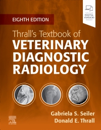
Thrall’s Textbook of Veterinary Diagnostic Radiology - Elsevier EBook on VitalSource, 8th Edition
by Gabriela Seiler, Dr.Med.Vet., DECVDI, DACVR and Donald E. Thrall, DVM, PhD
Elsevier eBook on VitalSource
ISBN:
9780323933803
Copyright:
2025
Publication Date:
09-20-2024
Page Count:
1024
Imprint:
Saunders
List Price:
$156.99
Most review copies are eBooks – how fast!
A review copy request is most likely to be fulfilled as an eBook on VitalSource rather than a print product, unless no eBook is available. eBooks become available in as little as a few hours. Print products will take between 7 and 10 days to arrive. To request a print copy, please contact us through the Evolve Support Center for further assistance or contact your Elsevier Sales Rep.
**Selected for 2026 Doody's Core Titles as an Essential Purchase in Veterinary Medicine**
Improve your students’ radiographic interpretation skills, regardless of their level of experience. Textbook of Veterinary Diagnostic Radiology, 8th Edition, is a one-stop resource covering the principles of radiographic technique and interpretation for dogs, cats, and horses. Within this bestselling text, high-quality radiographic images accompany clear coverage of diagnostic radiology, ultrasound, MRI, and CT. User-friendly direction helps students develop essential skills in patient positioning, radiographic technique and safety measures, normal and abnormal anatomy, radiographic viewing and interpretation, and alternative imaging modalities. This edition has been thoroughly revised to include the latest advances in the field, expand the number of image examples, and include a new ebook with every new print purchase!
Improve your students’ radiographic interpretation skills, regardless of their level of experience. Textbook of Veterinary Diagnostic Radiology, 8th Edition, is a one-stop resource covering the principles of radiographic technique and interpretation for dogs, cats, and horses. Within this bestselling text, high-quality radiographic images accompany clear coverage of diagnostic radiology, ultrasound, MRI, and CT. User-friendly direction helps students develop essential skills in patient positioning, radiographic technique and safety measures, normal and abnormal anatomy, radiographic viewing and interpretation, and alternative imaging modalities. This edition has been thoroughly revised to include the latest advances in the field, expand the number of image examples, and include a new ebook with every new print purchase!
-
- UPDATED! User-friendly content helps students develop essential skills in patient positioning, radiographic technique and safety measures, normal and abnormal anatomy, radiographic viewing and interpretation, and alternative imaging modalities
- NEW! The latest digital imaging information helps students stay up to date with the latest advances in the field
- NEW! An ebook version, included with every new print purchase, provides access to all the text, figures, and references, with the ability to search, customize content, make notes and highlights, and have content read aloud. Also included are videos, quizzes, and additional image examples of the most common diseases
- UPDATED! Current coverage of the principles of radiographic technique and interpretation for the most seen species in private veterinary practices and veterinary teaching hospitals includes the cat, dog, and horse
- Coverage of special imaging procedures such as the esophagram, upper GI examination, excretory urography, and cystography, helps in determining when and how these procedures are performed in today’s practice
- Content on abdominal ultrasound imaging helps in deciding on a diagnostic plan and interpreting common ultrasound findings
- An atlas of normal radiographic anatomy in each section makes it easier to recognize abnormal radiographic findings
- High-quality radiographic images clarify key concepts and interpretation principles
-
1. Radiation Protection and Physics of Diagnostic Radiology
2. Digital Radiographic Imaging
3. Physics of Ultrasound Imaging
4. Principles of Computed Tomography and Magnetic Resonance Imaging
5. Contrast Media in Diagnostic Imaging
6. Radiographic Interpretation
7. Errors and Pitfalls in Radiographic Interpretation
8. Radiographic Anatomy of the Axial Skeleton
9. Basic Principles of Radiographic Interpretation of the Axial Skeleton
10. Canine and Feline Dental Disease
11. The Skull and Nasal Cavities: Canine and Feline
12. Magnetic Resonance Imaging and Computed Tomographic Features of Brain Disease in Small Animals
13. The Equine Head
14. Radiography and Myelography of the Canine and Feline Vertebrae
15. Magnetic Resonance Imaging and Computed Tomography Features of Canine and Feline Spinal Cord Disease
16. Radiographic Anatomy of the Appendicular Skeleton
17. Principles of Radiographic Interpretation of the Appendicular Skeleton and Radiographic Features of Bone Tumors
18. Orthopedic Diseases of Young and Growing Dogs and Cats
19. Fracture Healing and Complications
20. Radiographic Signs of Joint Disease in Dogs and Cats
21. The Equine Stifle
22. Equine Tarsus
23. Equine Carpus
24. Equine Metacarpus and Metatarsus
25. Equine Fetlock Joint
26. Equine Pastern
27. Equine Foot
28. Principles of Radiographic Interpretation of the Thorax
29. Canine and Feline Larynx and Trachea
30. Pharynx, Upper Esophageal Sphincter, and Esophagus
31. Canine and Feline Thoracic Wall
32. Canine and Feline Diaphragm
33. Canine and Feline Mediastinum
34. Canine and Feline Pleural Space
35. Canine and Feline Cardiovascular System
36. Canine and Feline Lung
37. Equine Thorax
38. Principles of Radiographic Interpretation of the Abdomen
39. Peritoneal Space
40. Liver and Spleen
41. Kidneys and Ureters
42. Urinary Bladder and Urethra Chapter
43. Prostate Gland
44. Uterus, Ovaries, Vagina, and Testes
45. Stomach
46. Small Bowel
47. Large Bowel -
-
Ways of Reading
- The appearance of the text and page layout can be modified according to the capabilities of the reading system (font family and font size, spaces between paragraphs, sentences, words, and letters, as well as color of background and text)
- This e-publication is accessible to the full extent that the file format and types of content allow, on a specific reading device, by default, without necessarily including any additions such as textual descriptions of images or enhanced navigation
- No information about nonvisual reading is available
-
Conformance
- For detailed accessibility information, see Elsevier's website at https://www.elsevier.com/about/accessibility
- For queries regarding accessibility information, contact [email protected]
- The publication was certified on 18-11-2025
-
Navigation
- Table of contents to all chapters of the text via links
-
Rich Content
- No information is available
-
Hazards
- No information is available
-
Product Content
- No information is available
-
Legal Considerations
- No information is available
-
Additional Accessibility Information
- For readers with color vision deficiency, use of color (e.g., in diagrams, graphics and charts, in prompts, or on buttons inviting a response) is not the sole means of graphical distinction or of conveying information
- E-publication includes basic navigation (usually less detailed than TOC-based navigation)
- Where links, controls or buttons are included in the content, the purpose or functionality of each link, control or button is apparent from the associated text alone - or where it is unclear, separate link, control or button descriptions are provided
- All (or substantially all) textual matter is arranged in a single logical reading order (including text that is visually presented as separate from the main text flow, e.g., in boxouts, captions, tables, footnotes, endnotes, citations, etc.). Non-textual content is also linked from within this logical reading order. (Purely decorative non-text content can be ignored).
- The language of the text has been specified (e.g., via the HTML or XML lang attribute) to optimise text-to-speech (and other alternative renderings), both at the whole document level and, where appropriate, for individual words, phrases or passages in a different language.
-
Ways of Reading


Most review copies are eBooks – how fast!
A review copy request is most likely to be fulfilled as an eBook on VitalSource rather than a print product, unless no eBook is available. eBooks become available in as little as a few hours. Print products will take between 7 and 10 days to arrive. To request a print copy, please contact us through the Evolve Support Center for further assistance or contact your Elsevier Sales Rep.

 as described in our