ISBN:
9780443384462
Copyright:
2027
Publication Date:
02-25-2026
Page Count:
736
Imprint:
Elsevier
List Price:
$170.99

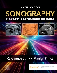
Sonography - Elsevier E-Book on VitalSource, 6th Edition
by Reva Curry and Marilyn Prince
Elsevier eBook on VitalSource
ISBN:
9780443384462
Copyright:
2027
Publication Date:
02-25-2026
Page Count:
736
Imprint:
Elsevier
List Price:
$170.99
Most review copies are eBooks – how fast!
A review copy request is most likely to be fulfilled as an eBook on VitalSource rather than a print product, unless no eBook is available. eBooks become available in as little as a few hours. Print products will take between 7 and 10 days to arrive. To request a print copy, please contact us through the Evolve Support Center for further assistance or contact your Elsevier Sales Rep.
Help students learn how to accurately identify abnormalities in ultrasound images! Sonography: Introduction to Normal Structure and Function, 6th Edition, grounds students in what normal anatomy and physiology look like from an ultrasound perspective, enabling them to better recognize abnormalities. Each chapter begins with coverage of anatomy and physiology followed by a wealth of ultrasound images to visually reinforce concepts and increase students’ comfort with normal anatomy as it appears during scanning. A consistent chapter format also makes the content easy to navigate and reinforces standard protocols for scanning each area of the body.
-
- UPDATED! 100+ images and accompanying illustrations visually clarify key concepts and give you a better picture of what you should see in scans
- NEW! AI-empowered ultrasonography coverage explores the emergence of this technology and its impact on clinical practice
- NEW! Targeted callouts aligned with ARDMS registry content enhance exam preparation
-
- UPDATED! 100+ images and accompanying illustrations visually clarify key concepts and give students a better picture of what they will see in scans
- NEW! AI-empowered ultrasonography coverage explores the emergence of this technology and its impact on clinical practice
- NEW! Targeted student callouts aligned with ARDMS registry content enhance exam preparation
- Detailed line drawings accompany most sonograms, reinforcing what you should notice on the scan and offering confirmation if you are uncertain
- Highly visual content leads with images and uses narrative to support those visuals
- Quality control protocol information helps you recreate the most optimal scanning settings and techniques
- Reference charts offer relevant information for types of sonography, including associated physicians, common diagnostic tests, laboratory values, vasculature, and affecting chemicals
-
-

-
 Workbook and Lab Manual for Sonography - Elsevier E-Book on VitalSourceElsevier eBook on VitalSource
 -
 Workbook and Lab Manual for SonographyPaperback
 -
 SonographyHardcover
-
-
SECTION I CLINICAL APPLICATIONS
1. Before, During, and After the Ultrasound Examination
2. Ultrasound Instrumentation “Knobology,” Imaging Processing, and Storage
3. General Patient Care
4. Introduction to Ergonomics and Sonographer Safety
SECTION II SONOGRAPHIC APPROACH TO UNDERSTANDING ANATOMY
5. Interdependent Body Systems
6. Anatomy Layering and Sectional Anatomy
7. Embryology
8. Introduction to Laboratory Values
SECTION III ABDOMINAL SONOGRAPHY
9. The Abdominal Aorta
10. The Inferior Vena Cava
11. The Portal Venous System
12. The Liver
13. The Biliary System
14. The Pancreas
15. The Urinary System and Adrenal Glands
16. Abdominal Vasculature Flow Dynamics
17. The Spleen
18. The Gastrointestinal System
SECTION IV PELVIC SONOGRAPHY
19. The Male Pelvis: Prostate Gland and Seminal Vesicles Sonography
20. The Female Pelvis
SECTION V OBSTETRICAL AND NEONATAL SONOGRAPHY
21. First-Trimester Obstetrics (0–12 Weeks)
22. Second and Third Trimester Obstetrics (13–42 Weeks)
23. High-Risk Obstetric Sonography
24. Fetal Echocardiography
25. The Neonatal Brain
SECTION VI SMALL PARTS SONOGRAPHY
26. The Thyroid and Parathyroid Glands
27. Breast Sonography
28. Scrotal and Penile Sonography
SECTION VII SPECIALTY SONOGRAPHY
29. Pediatric Echocardiography
30. Adult Echocardiography
31. Vascular Technology: Carotid Artery and Peripheral Venous Duplex
SECTION VIII ADVANCES IN SONOGRAPHY
32. 3D/4D/5D Sonography
33. Interventional and Intraoperative Ultrasound
34. Musculoskeletal Sonography
35. Pediatric Sonography -
-
Ways of Reading
- The appearance of the text and page layout can be modified according to the capabilities of the reading system (font family and font size, spaces between paragraphs, sentences, words, and letters, as well as color of background and text)
- This e-publication is accessible to the full extent that the file format and types of content allow, on a specific reading device, by default, without necessarily including any additions such as textual descriptions of images or enhanced navigation
- All content can be read as read aloud speech or dynamic braille
- Has alternative text descriptions for images
-
Conformance
- The certifier's credential is https://bornaccessible.benetech.org/certified-publishers/
- For detailed accessibility information, see Elsevier's website at https://www.elsevier.com/about/accessibility
- For queries regarding accessibility information, contact [email protected]
- The publication was certified on 24-12-2025
-
Navigation
- Table of contents to all chapters of the text via links
- Index with links to referenced entries
- Elements such as headings, tables, etc. for structured navigation
- Page list to go to pages from the print source version
-
Rich Content
- Math formulas in accessible format (MathML)
- Information-rich images are described by extended descriptions
-
Hazards
- The publication contains no hazards
-
Product Content
- Content includes any type of illustrations.
- The primary content is text.
- Content includes maps and/or other cartographic content.
- Content includes a significant number of actionable (clickable) web links to external content, downloadable resources, supplementary material, etc.
- Content includes a significant number of actionable (clickable) cross-references, hyperlinked notes and annotations, or with other actionable links between largely textual elements (e.g., quiz/test questions, 'choose your own ending', etc.).
- Content includes photographs, whether in a plate section / insert or not.
- Content includes figures, diagrams, charts and/or graphs, including other 'mechanical' (i.e. non-photographic) illustrations.
- Content includes images displayed in a specific order for the purpose of graphic storytelling or giving information (e.g. graphic novels, comics and manga). May include text integrated into the image (as speech and thought bubbles, textual 'sound' effects, captions etc.).
- Content includes text within images, including text-as-text embedded in diagrams, charts, or within images containing speech balloons, thought bubbles, captions, etc.
- Content includes visual content that is purely decorative and is not necessary to understanding of the content.
- Content includes mathematical notations, formulae.
- At least some text - including text within other images - is 'text as an image' (i.e., a picture of text).
-
Legal Considerations
- No information is available
-
Additional Accessibility Information
- Content is enhanced with ARIA roles to optimize organization and facilitate navigation
- Page breaks included from the original print source
- For readers with color vision deficiency, use of color (e.g., in diagrams, graphics and charts, in prompts, or on buttons inviting a response) is not the sole means of graphical distinction or of conveying information
- The body text is presented with a contrast ratio of at least 4.5:1 (or 3:1 for large/heading text)
- Ultra-high contrast between text and background
- E-publication includes basic navigation (usually less detailed than TOC-based navigation)
- Where links, controls or buttons are included in the content, the purpose or functionality of each link, control or button is apparent from the associated text alone - or where it is unclear, separate link, control or button descriptions are provided
- All (or substantially all) textual matter is arranged in a single logical reading order (including text that is visually presented as separate from the main text flow, e.g., in boxouts, captions, tables, footnotes, endnotes, citations, etc.). Non-textual content is also linked from within this logical reading order. (Purely decorative non-text content can be ignored).
- Content provides explanations for unusual words, abbreviations, acronyms, idioms, jargon in an accessible form, such as glossaries, scripted pop-ups.
- Where interactive content is included in the product, controls are provided (e.g., for speed, pause and resume, reset) and labelled to make their use clear.
- The language of the text has been specified (e.g., via the HTML or XML lang attribute) to optimise text-to-speech (and other alternative renderings), both at the whole document level and, where appropriate, for individual words, phrases or passages in a different language.
-
Ways of Reading
Most review copies are eBooks – how fast!
A review copy request is most likely to be fulfilled as an eBook on VitalSource rather than a print product, unless no eBook is available. eBooks become available in as little as a few hours. Print products will take between 7 and 10 days to arrive. To request a print copy, please contact us through the Evolve Support Center for further assistance or contact your Elsevier Sales Rep.

 as described in our