ISBN:
9781437704150
Copyright:
2014
Publication Date:
05-28-2014
Page Count:
640
Imprint:
Saunders
List Price:
$209.99
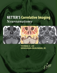
Netter’s Correlative Imaging: Neuroanatomy, 1st Edition
by Thomas C. Lee, MD and Srinivasan Mukundan, MD, PhD
Hardcover
ISBN:
9781437704150
Copyright:
2014
Publication Date:
05-28-2014
Page Count:
640
Imprint:
Saunders
List Price:
$209.99
In Stock
This item has low stock levels and may be back-ordered. We'll let you know if it is back-ordered, and you will not be charged until the item ships.
-
- View the brain, spinal cord, and cranial nerves, as well as head and neck anatomy through modern imaging techniques in a variety of planes, complemented with a detailed illustration of each slice done in the instructional and aesthetic Netter style.
- Find anatomical landmarks quickly and easily through comprehensive labeling and concise text highlighting key points related to the illustration and image pairings.
- Correlate patient data to idealized normal anatomy, always in the same view with the same labeling system.
-
PART 1 - BRAIN
1 - Overview of Brain
2 - Brain
3 - Thalamus and Basal Ganglia
4 - Limbic System
5 - Brainstem and Cranial Nerves
6 - Ventricles and Cerebrospinal Fluid Cisterns
7 - Sella Turcica
PART 2 - HEAD AND NECK
8 - Overview of Head and Neck
9 - Paranasal Sinuses
10 - Orbits
11 - Mandible and Muscles of Mastication
12 - Temporal Bone (Middle Ear, Cochlea, Vestibular System)
13 - Oral Cavity, Pharynx, and Suprahyoid Neck
14 - Hypopharynx, Larynx, and Infrahyoid Neck
PART 3 - SPINE
15 - Overview of Spine
16 - Spine
-
No accessibility information is available for this product.
 as described in our