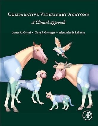
Comparative Veterinary Anatomy: A Clinical Approach describes the comprehensive, clinical application of anatomy for veterinarians, veterinary students, allied health professionals and undergraduate students majoring in biology and zoology. The book covers the applied anatomy of dogs, cats, horses, cows and other farm animals, with a short section on avian/exotics, with a focus on specific clinical anatomical topics.

Q: What is unique about this veterinary anatomy book?
A: This book links the basic anatomy course with the essential applied anatomy needed in holistically evaluating, diagnosing, and treating multiple veterinary species.
Q: Why is this book important for first year veterinary students?
A: First-year veterinary students will learn the significance of basic and applied anatomy from the beginning of their professional education. This text strives to demonstrate the clinical application of anatomy and its terminology, along with how it relates to diagnostics, such as imaging and therapeutics, and surgery, along with the many nuances in developing into a well-rounded clinician.
Q: Do students in their second, third, and fourth years of veterinary school benefit from having this book in clinics?
A: Every course in medicine, surgery, reproduction, and various imaging studies relies on clinically oriented anatomy at all levels of study. The book will be a lifelong career reference in all personal and academic libraries.
Q: Who else might benefit from keeping this book on hand?
A: Veterinarians, veterinary nurses and technicians, surgeons, emergency clinicians, internal medicine, theriogenology, imaging specialists, comparative anatomists, graduate students in clinical related subjects, undergraduate students majoring in the biological and zoological sciences, anatomy and clinical faculty, allied health professionals, and anyone with an interest in animal science and veterinary medicine.
Q: What are some of the best features of this book?
A: The book makes anatomy clinically relevant across species with a comparative focus. It covers the primary veterinary species seen in practice; contains layered landscape color figures at the beginning of each section emphasizing the specific species from skeleton to skin; provides anatomically accurate color figures for each anatomical region studied; and correlates gross anatomy with diagnostic imaging findings such as digital radiology, ultrasound, CT, MRI, nuclear scintigraphy, and PET. The book facilitates both independent and classroom learning, as well as being a ready reference resource.
Q: Who are the contributing authors, and how were they chosen?
A: Section editors and contributing authors are world-renowned clinicians and clinical anatomists in their specialty and species. We were honored to have the late Dr. de Lahunta, a world-renowned anatomist, oversee the book’s production.